Home
/ Anatomy Of Ribs And Muscles, Rib Wikipedia, Jul 16, 2019 · small muscles running between the ribs, known as the external intercostal muscles, lift the ribs during deep breathing to further expand the chest and lungs and provide even more air to the body.
Anatomy Of Ribs And Muscles, Rib Wikipedia, Jul 16, 2019 · small muscles running between the ribs, known as the external intercostal muscles, lift the ribs during deep breathing to further expand the chest and lungs and provide even more air to the body.
Anatomy Of Ribs And Muscles, Rib Wikipedia, Jul 16, 2019 · small muscles running between the ribs, known as the external intercostal muscles, lift the ribs during deep breathing to further expand the chest and lungs and provide even more air to the body.. These include the erector spinae muscle group and posterior serratus muscles on the back, and the abdominal wall muscles on the front. Rib below its origin, medial to the angle: Lying exposed between the protective bones of the superiorly located ribs and the inferiorly located pelvic girdle, the muscles of this region play a critical role in protecting the. Serratus posterio r consists of two muscles that assist respiration; Its function is to elevate the ribs.
Its primary action is to extend, adduct and internally rotate the upper arm. The upper edge is round and the lower sharp. Human muscles · april 17, 2020. The part of the muscle is thought to depress the ribs. It is one of the borders of the superior thoracic aperture.
Anatomy Of Rib Muscles Anatomy Drawing Diagram from i.pinimg.com The inferior or lower border of each rib 5 insertion: Rib below its origin, medial to the angle: Others attach indirectly because they are attached to the cartilage of the rib above. Choose from 500 different sets of anatomy flashcards on quizlet. The superior or upper border of the rib below 5 the muscle fibers run slantingly, in an inward (toward the center of the body) and downward direction 2, connecting the 12 ribs to each other.the external intercostal muscles form the external intercostal membrane that extends to attach to the sternum 6. Its primary action is to extend, adduct and internally rotate the upper arm. The part of the muscle is thought to depress the ribs. The thoracic wall is formed by 12 ribs, 12 thoracic vertebrae, cartilage, sternum, and five muscles.
There are three layers of intercostal muscles:
Flexes, laterally flexes, or rotates vertebral column to same side. Our ribcage exists to protect the heart and lungs. The thoracic wall is formed by 12 ribs, 12 thoracic vertebrae, cartilage, sternum, and five muscles. Choose from 500 different sets of anatomy flashcards on quizlet. The first seven ribs attach directly to the sternum through cartilage that forms at the end of each rib. At the chest, many rib bones connect to the sternum via costal cartilage,. Between each rib lie several layers of intercostal muscles that are responsible for expanding and shrinking. Serratus posterio r consists of two muscles that assist respiration; The upper edge is round and the lower sharp. It is one of the borders of the superior thoracic aperture. The rectus abdominis runs between the ribs and the pubic bone and supports movements between the rib cage and the pelvis. The serratus anterior (magnus) muscle (fig. Human anatomy drawing human figure drawing anatomy study anatomy art anatomy reference figure drawing reference pose reference anatomy bones body anatomy.
It is one of the borders of the superior thoracic aperture. The sections that follow trace the grades and quality, the origins, anatomy and cuts, the cooking styles, and the flavors of meat. Our ribcage exists to protect the heart and lungs. The more anterior your cut, the more spinalis you'll find in the steak. This part of the muscle is thought to elevate the ribs.
Pin On Thoracic from i.pinimg.com They help stabilize your upper body and help you breathe. These are fairly small and insignificant muscles: It functions in movement, respiration, and protection of the thoracic cavity. Complete anatomy version 2.3.7 (4120), 2017 (1) the overlying muscles in the thoracic region span across multiple ribs and vertebrae, helping to generate force and movement. The more anterior your cut, the more spinalis you'll find in the steak. It is one of the borders of the superior thoracic aperture. They comprise of thin slips of muscle, which run from the internal surface of one rib, to second and third ribs below. Ribs 3 to 9 are considered typical ribs and have.
The serratus anterior (magnus) muscle (fig.
These are fairly small and insignificant muscles: It functions in movement, respiration, and protection of the thoracic cavity. Jul 16, 2019 · small muscles running between the ribs, known as the external intercostal muscles, lift the ribs during deep breathing to further expand the chest and lungs and provide even more air to the body. Human anatomy drawing human figure drawing anatomy study anatomy art anatomy reference figure drawing reference pose reference anatomy bones body anatomy. At the chest, many rib bones connect to the sternum via costal cartilage,. The part of the muscle is thought to depress the ribs. It is one of the borders of the superior thoracic aperture. Thoracolumbar fascia, inguinal ligament, and iliac crest. The superior fibres originate from the spinous processes of the c7 to t3 vertebrae and attach to the superior borders of ribs two to four. Deep cervical a., intercostal aa. The first seven ribs attach directly to the sternum through cartilage that forms at the end of each rib. With the upper ribs, closer to the nodule (and in the case of lower ribs, a little further from the nodule) they are curved and have a rough surface that connects them with muscles, angulus costae. The muscles of the abdomen, lower back, and pelvis are separated from those of the chest by the muscular wall of the diaphragm, the critical breathing muscle.
Изображение rib cage muscles diagram. The muscles of the abdomen, lower back, and pelvis are separated from those of the chest by the muscular wall of the diaphragm, the critical breathing muscle. The superior or upper border of the rib below 5 the muscle fibers run slantingly, in an inward (toward the center of the body) and downward direction 2, connecting the 12 ribs to each other.the external intercostal muscles form the external intercostal membrane that extends to attach to the sternum 6. Choose from 500 different sets of anatomy flashcards on quizlet. Related posts of muscle anatomy ribs muscle anatomy posterior.
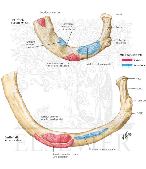
Muscle Attachments Of Ribs from www.netterimages.com Related posts of muscle anatomy ribs muscle anatomy posterior. It is one of the borders of the superior thoracic aperture. The upper edge is round and the lower sharp. Human muscles · april 17, 2020. The superior or upper border of the rib below 5 the muscle fibers run slantingly, in an inward (toward the center of the body) and downward direction 2, connecting the 12 ribs to each other.the external intercostal muscles form the external intercostal membrane that extends to attach to the sternum 6. Ribs 3 to 9 are considered typical ribs and have. Our ribcage exists to protect the heart and lungs. Jul 16, 2019 · small muscles running between the ribs, known as the external intercostal muscles, lift the ribs during deep breathing to further expand the chest and lungs and provide even more air to the body.
There are three layers of intercostal muscles:
The rectus abdominis runs between the ribs and the pubic bone and supports movements between the rib cage and the pelvis. The inferior or lower border of each rib 5 insertion: The thoracic vertebral bodies and intervertebral discs make the posterior thoracic wall. Serratus posterio r consists of two muscles that assist respiration; Ribs 3 to 9 are considered typical ribs and have. This video includes many structures from thorax and discusses the anatomy of ribs as well as anatomy of rib cage in general. Its function is to elevate the ribs. Rib below its origin, medial to the angle: The functions of the ribs are critical, as they protect the contents of the thoracic cavity and mediastinum, move superiorly, inferiorly, anteriorly and posteriorly to facilitate breathing, provide a place where some muscles originate or attach, and play a role in erythropoiesis during development. Click card to see definition 👆. The first seven ribs attach directly to the sternum through cartilage that forms at the end of each rib. Human anatomy drawing human figure drawing anatomy study anatomy art anatomy reference figure drawing reference pose reference anatomy bones body anatomy. Rib eye steaks are mainly composed of the longissimus dorsi muscle (the eye portion of the steak) and the spinalis dorsi muscle.
These are fairly small and insignificant muscles: anatomy of ribs. Click card to see definition 👆.